Posts

Showing posts with the label Case report

Caries detector dye

Image
 I was going to make a post on caries detector dye because I find it to be such a useful aid during restorative procedures. Then I realised I had already made a post about in 2020. See below. https://dental-tidbits.blogspot.com/2020/09/a-note-on-caries-detector-dye.html  I pretty much agree with my thoughts on the previous post. At the time in 2020 I was using the dye fairly regularly as they had it at the public clinic I was at. I think my use dropped off as I transitioned to private practice and not every clinic I worked at had access to it. Now if I worked at a clinic without access to caries detector dye it would be something I would be requesting or purchasing myself.  Not that I need it to practice, no one needs it, but using it regularly will assist in consistency of your restorations. For proper adhesion to tooth structure, you must ensure that you are bonding to sound enamel and dentine. Therefore caries must be removed to an acceptable standard, clean margins ar...

When to reline/modify vs when to remake a denture

Image
For some reason, I had a long period of no denture cases coming in. For maybe 1-2 years I did a couple of fractured denture repairs but no new denture cases. Lately there have been some new ones coming in which have been enjoyable to work with. I thought I would make a post regarding modification vs remaking of dentures. Of course by modification I don't mean something simple like adjusting a denture sore spot, where we obviously wouldn't remake a denture, but for where there is a significant discrepancy between what the denture should be like and what it is like e.g changes to the fit or teeth. One common dilemma when a patient comes in with an old set of dentures is do we do nothing, do we try to improve the current denture, or do we start again and create a new set for the patient. My take on this is simple, if I can make an improvement to their current denture and this improvement will make a significant difference to the patient then it is justifiable to do something for t...

Review of an old blog post

Today I had the opportunity to review a previous case: http://dental-tidbits.blogspot.com/2018/08/getting-good-contact-with-large-gics.html http://dental-tidbits.blogspot.com/2019/05/review-of-alternative-technique-for-gics.html The entire GIC debonded a couple of weeks ago about a 11 months after placement when the patient was biting on a mintie. It's not a bad result but still needs management. The idea of the initial GIC placement was as a temporary to monitor the pulp status and the tooth has been otherwise asymptomatic. One might question if it's asymptoamtic due to a receeded pulp or a necrotic pulp. The failure was adhesive with the entire GIC lost save for the vitrebond liner so this was placed back when I was doing more indirect pulp caps. As enough time had passed for pulp review I replaced the restoration in composite. I'm a lot less worried these days about causing pain with wedges. The patient consented to treatment without LA and understands there will ...

6 month review of cracked tooth

Review of case 6 months ago: http://dental-tidbits.blogspot.com/2018/12/case-report-cracked-tooth.html The lower 6 remains asymptomatic but the upper 6 has become symptomatic with cracked tooth syndrome. The other dentist in the practice has seen her and will perfom cuspal coverage of this tooth. On followup if I were seeing her I would consider taking follow up photos, pulp test and a PA radiograph. Muscles of mastication are painful to tenderness. She is likely a bruxer due to the significant wear on the 7, multiple cracks and pain in her muscles. The sign of heavy occlusion on the 7 is also a good sign that she may have her MIP anterior to her CR postition. Seating of the condyles into CR will result in posterior traction of the mandible and a slight separation of the anterior teeth. This tends to bring the most posterior tooth i.e the lower 7s into heavy contact. This can be reproduced by adding leaves in a leaf gauge to the anterior teeth and checking the occlusion of the pos...

Review of alternative technique for GICs

A while back, I explored a technique idea for getting stronger contacts with GICs: http://dental-tidbits.blogspot.com/2018/08/getting-good-contact-with-large-gics.html Thinking back on this technique, I don't think it's a good idea because the premise involves compacting the GIC after the gel stage i.e when there is some solidity to the material and it has lost it's sheen. Prof. Ian Meyers maintains that the best bond to tooth structure will be if the GIC is placed against the tooth while it is still flowy and shiny however this method involves manipulation of the material after this stage. Unfortunately, even light compaction of the GIC is likely to cause movement of the material away from the tooth that will lower or even completely remove its bond strength. This is likely why there was a restoration fracture soon after placement. Alternative techniques would be to place a wooden wedge with or without a V ring to get a very tight contact initially with the GIC. If t...
Today I had an interesting occurrence whereby there was a sinus infection associated with a dental infection. While I was going through routine consent, he responded to the risk of OAC by saying he has had recurrent sinus infections in the past. he has apparently seen his GP who found nasal polyps and has recurrent sinus infections that he is prescribed antibiotics for. On a side note it is important not to take everything the patient reports as a fact. One would hope that the GP offered more investigation i.e referral to an ENT rather than symptomatic relief from antibiotics. Who knows, maybe the patient was offered this but declined or maybe he sees a different GP every time. Additionally, on the PA it did seem as though the palatal root was close to the maxillary sinus with a periapical radiolucency involved. These should be warning signs to get prooper consent form the patient and inform them of the possible adverse outcomes of treatment and the possible follow up strategies includ...

Case report- Cracked tooth

Image
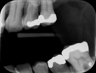
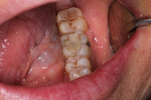
The following is a case report of a procedure where in hindsight I would do things a little differently This patient is a 50 Year old female who presented with the occasional pain to bite down on a lower left tooth. Immediately when I hear this I have an idea in my head as to the possible causes. Firstly the location seems to be fairly reproducible and it only appears to happen when she bites on a certain tooth. This almost completely rules out myofascial pain which would be more constant and deep and would be sore if she bit on any tooth on the affected side. Palpation of the masseter and lateral pterygoid will assist in ruling this out. Do be careful as there is often a secondary myofascial pain from the toothache or from bruxers who often have cracked teeth as a finding. Acute periapical pathology is an option but this would often lead to extended pain after biting and clinical examination should reveal tenderness to percussion which is reproducible. Pulp testing assists in determ...

Getting a good contact with large GICs

Image
Today I was in a situation where there was a 46 tooth with an MODB fracture. The mesial and distal margins were equigingval. The tooth is set for an endo or and exo but I did not have the required equipment to perform either (In the dental truck). So the plan is a temporary restoration and the patient will seek treatment privately. There are two options from here. -Restore to decent contour and contact -Restore with a stump with open contacts Ideally I would have a good contact to stop food packing but at a copmromise I would consider a wide open contact to allow interdental brushes to fit in. Without anaesthesia which was my plan, Wedges would be painful and making a contact would be difficult. This was due to the equigingival margin. In the past to make a decent contact I have used a light cure GIC like Fuji2LC and restored in stages. Sectional matricies are usually too unstable to be used due to the minimal surface area they are in contact with the tooth and there is the same ...

Cracks, cracks, cracks

Image
Today I had a case that taught me not to focus on the obvious cause of pain. A patient came in complaining of pain on the 25. the 24 had previously been filled by another dentist and I assume it must have been a carious exposure. Checking the xrays before the patient arrived I settled on the 24 as the probably cause of pain. She presented and was very sure that it was the 25 painful to hot and cold. There was an MOD amalgam in the 25 that appeared sound. I still thought it was the 24... Cold test was very painful for both but more so for the 25. The curiosity was that there was an exaggerated response to pain on the 25 as it appeared sound. I was very ready to extract the 24, so much so that the needle was right near the tooth for LA. I decided to test the teeth once more and again a more exaggerated response from the 25. Still unsure if the patient was confused, I did a heat test for the first time. Rubber dam isolation of the 25 and washing of the tooth with warm water from a monoje...