Posts

Showing posts with the label cracked tooth

Cracked tooth- Metal Band tips

 Today I had to cement a metal band around a virgin lower 6 with a distal crack into the pulp. Difficulties I've had in the past with metal bands were:  -    Band selection: Today I used a periodontal probe to measure the mesial-distal dimension of the tooth. The issue with the probe I had was the measurements didn't go far enough to measure the whole tooth in one go so I had to guess the last few millimetres. ensure you use a proble that has measurements far along enough to measure the entire width of the tooth. The mesiodistal width is the most important measurements to fit snugly as the width of the band is often limited by the position of the adjacent teeth. Ensure you measure from the widest part of the tooth not the marginal ridge as the band has to fit all the way past where the contact point is. The more worn the occlusal surface of the tooth the closer the occlusal table dimension will resemble the widest part of the tooth. With a tooth that isn't worn signi...
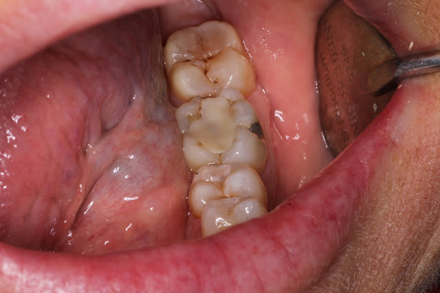
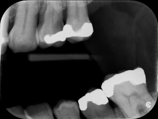
6 month review of cracked tooth

Review of case 6 months ago: http://dental-tidbits.blogspot.com/2018/12/case-report-cracked-tooth.html The lower 6 remains asymptomatic but the upper 6 has become symptomatic with cracked tooth syndrome. The other dentist in the practice has seen her and will perfom cuspal coverage of this tooth. On followup if I were seeing her I would consider taking follow up photos, pulp test and a PA radiograph. Muscles of mastication are painful to tenderness. She is likely a bruxer due to the significant wear on the 7, multiple cracks and pain in her muscles. The sign of heavy occlusion on the 7 is also a good sign that she may have her MIP anterior to her CR postition. Seating of the condyles into CR will result in posterior traction of the mandible and a slight separation of the anterior teeth. This tends to bring the most posterior tooth i.e the lower 7s into heavy contact. This can be reproduced by adding leaves in a leaf gauge to the anterior teeth and checking the occlusion of the pos...

Presentation of cracks

When examining teeth, apart from from caries, restorations and the periodontal condition, cracks are a common finding, more so the older the patient is. Common places that cracks are seen are the mesial and distal marginal ridges of posterior teeth as well as the mid buccal and lingual grooves. Craze lines are often seen on the labial surface of anterior teeth. Cracks are most commonly seen on mandibular molars, then upper premolars. Teeth more posterior are closer to the muscles of mastication and to the fulcrum point of the TMJ therefore forces tend to be higher on these teeth. Upper molars tend to erupt tipping towards the buccal and the palatal cusps as a result protrude occlusally. These plunger cusps tend to interfere with the lower molars cusps on lateral excursion and predispose to cracks. When a crack is detected, carefully view the level of the posterior occlusal plane in a mouth mirror from the palatal aspect. You should be able to see if a plunger cusp sits above the le...

Case report- Cracked tooth

Image
The following is a case report of a procedure where in hindsight I would do things a little differently This patient is a 50 Year old female who presented with the occasional pain to bite down on a lower left tooth. Immediately when I hear this I have an idea in my head as to the possible causes. Firstly the location seems to be fairly reproducible and it only appears to happen when she bites on a certain tooth. This almost completely rules out myofascial pain which would be more constant and deep and would be sore if she bit on any tooth on the affected side. Palpation of the masseter and lateral pterygoid will assist in ruling this out. Do be careful as there is often a secondary myofascial pain from the toothache or from bruxers who often have cracked teeth as a finding. Acute periapical pathology is an option but this would often lead to extended pain after biting and clinical examination should reveal tenderness to percussion which is reproducible. Pulp testing assists in determ...

Cracks, cracks, cracks

Image
Today I had a case that taught me not to focus on the obvious cause of pain. A patient came in complaining of pain on the 25. the 24 had previously been filled by another dentist and I assume it must have been a carious exposure. Checking the xrays before the patient arrived I settled on the 24 as the probably cause of pain. She presented and was very sure that it was the 25 painful to hot and cold. There was an MOD amalgam in the 25 that appeared sound. I still thought it was the 24... Cold test was very painful for both but more so for the 25. The curiosity was that there was an exaggerated response to pain on the 25 as it appeared sound. I was very ready to extract the 24, so much so that the needle was right near the tooth for LA. I decided to test the teeth once more and again a more exaggerated response from the 25. Still unsure if the patient was confused, I did a heat test for the first time. Rubber dam isolation of the 25 and washing of the tooth with warm water from a monoje...

ADA patient information sheet series- Cracked tooth syndrome

I've picked up some patient information sheets from work, just going to spend a few posts summarising the key points of these. -Usually occurs on a molar or premolar (in order of likelihood): 1. Upper premolar 2. Lower molar 3. Upper molar 4. Lower premolar Symptoms: -Sharp and erratic pain on chewing or after release of biting pressure. But not all cracks cause pain  -Pain or discomfort when exposed to cold or hot liquids or food -Sensitivity to sweet -Difficulty in pinpointing location of pain -If the crack extends blow the gum, a periodontal pocket may be present -Often a history of other cracked teeth Causes: -More likely in recent times as teeth are retained for longer with larger restorative cycles and teeth more prone to fracture -Stress leading to grinding of teeth especially at night -Time, wear and tear from chewing, grinding and clenching -Chewing on hard foods e.g ice, sweets or pencils -Trauma especially if upper and lower teeth have been rammed to...
When looking at out of place dental radiolucencies, check for enamel abnormalities e.g enamel fractures that may be on the strangest places and can appear to be carious on the x ray. Radiolucent areas under restorations may be voids or bases. Track cracks with your probe. Separation indicates a need for intervention and if they track subgingivally on the most posterior tooth this requires referral to an endodontist to assess the tooth for suitability for restoration.

More on cracks

Observed at DrDs today and he reinforced the need to check teeth for cracks. He pointed out cracks on teeth and asked" what should we do about this?" The general answer was nothing (if there was no symptoms). But when there is a cracks in a symptomatic tooth then we are at a loss of what to do. He removed the restoration and placed a GIC temporary. He had the choice of placing a corticosteroid medicament over the base of the prep to sedate the pulp but that would just mask the symptoms and we wouldn't know if the disappearance of symptoms were due to replacing the restoration or due to the corticosteroid and we would not be sure of the state of the pulp. If the pulp was healthy on review, restoration or crown is the answer. If it is non vital, RCT then crown or exo is the answer. He also advised that if there were symptomstic cracks on an unrestored tooth. crowning asap is necessary.

More on cracks- Caries

Cracks allow bacteria to enter down the passageway and cause caries in the tooth. We often look for signs of caries radiographically and look for a lesion in enamel. Deep dentine caries can be present without enamel shadowing as the caries process starts through a crack which is rarely visible radiographically

Cracks in teeth

Cracks are practically ubiquitous and with careful searching cracks can be found in almost any patient. Be wary of patients who present with unexplained sensitivity, heavily restored teeth, pain on biting and release of pressure. Cracks can still appear in unrestored teeth and fractures associated with these are generally much worse than in restored teeth and indicaate excessive forces are being generated e.g bruxism. Crowning is not always the way to go for any crack and although cracks are undiagnosed, they are also overtreated. The amount and extent of cracks should be matched up to the patients age e.g excessive cracks on young patients may require intervention. Also strategic teeth may consider early intervention e.g lonely molars with large restorations to "protect those teeth long term".  Treatment doesn't specify crowning teeth but involves cusp coverage which may be in the form of a porcelain overlay