A case study with some lessons
This is a string of photos I took almost two years ago and have been meaning to write about for a while. There were a few things I did wrong and a few lessons I have taken from the case. My memories are a bit sketchy on the exact details of the case but I will do the best I can.
-The patient presented to me complaining initially of spontaneous pain to the lower left second molar exacerbated by temperature changes. To me that is sounding like a pulpitis of some sort, leaning towards the irreversible diagnosis clinically.The 37 tooth had a couple of deficiencies on the occlusal and lingual aspects as well as an occlusal amalgam and a mesial crack apparent.
-As there was no visible carious lesion I chalked the problem down to the deficiencies in the tooth and patched them up with GIC. I didn't investigate thoroughly into the diagnosis, didn't perform pulp tests or even take an Xray. I am unsure why I didn't do this but possibly it was that the patient was very anxious and I wanted to do the minimum possible on her as it was the first visit and at the time I was always hesitant to perform treatment too invasive on these patients. Now I realise that it is important to accept that patients will be uncomfortable to an extent and it is more detrimental to perform an inadequate diagnosis leading to treatment that may not be effective.
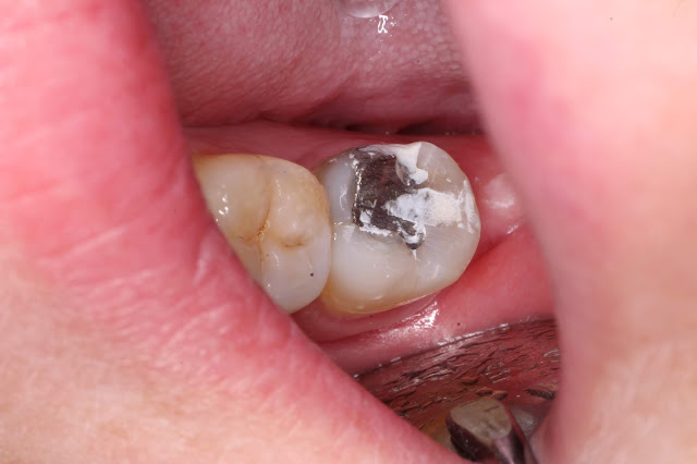
-I took the photo below at the end of that appointment. You can see the recently set GIC, retained occlusal amalgam and the mesial crack which I am not sure was apparent to me at the time. Even without an xray you can assume that the amalgam is quite deep as it takes up about a quarter of the tooth on the surface and the spread of caries at the time of placement was unlikely to be shallow. The crack is apparent due to the shade change of the mesiolingual cusp and we would be concerned about cuspal fracture just due to the loss of structural integrity of the cusp. Definitely in the future I would consider using a frac finder, pulp testing and taking a radiograph. What I wouldn't have seen at the time was the large wear facet on the distal aspect of the occlusal surface. These days I would definitely be pulling out a leaf gauge and checking for the contacts in centric relation as well as checking the alignment of the opposing teeth to check for a plunging cusp.
- The patient returned for another appointment with the tooth still in pain. The mesiolingual cusp had fractured off and it was apparent that there was mesial caries that had contributed along with the large restoration to undermining the cusp. I don't think I took a radiograph here and I am unsure why but I don't have any on record. Possibly at this point I assumed that the pain was from the crack and now the fracture had occured, the problem was solved. I anaesthetised the patient, placed a rubber dam and removed the restorations and caries to investigate. On the photo below you can just see my GIC adjacent to the clamp on the lingual aspect. I am sure that the deficiency on the lingual aspect at initial presentation was a thin piece of enamel that had cracks running through it. After that came off then the mesiolingual cusp was the next weakest part. On this photo you can see the good example of wear facets on the mesiobuccal and distobuccal cusps as well as the shiny wear facet on the inclined plane on the amalgam adjacent to the mesiolingual cusp site. Last visit I didn't use any rotary instruments when placing the GIC so I am certain this was due to occlusal function.
-I removed the old restoration, cleared the caries on the mesial but noted that there was caries underneath the amalgam restoration. This is light brown so I assume has developed fairly recently and is quickly progressing. I stopped to take a photo at this point partly for communication with the patient and partly because I didn't have a radiograph proving there was caries underneath this restoration.
-I excavated the caries and noted that there was a carious exposure of the pulp. It was red but not bleeding which indicated that this part of the pulp was non vital but very recently so. I can't recall the details of what I did here but I did temporise the tooth and finally took a PA radiograph. It shows that the temporary restoration extended right up and into the mesial pulp horns and there was a furcal radiolucency. It doesn't look as though I opened up the pulp chamber or placed any medicaments in the pulp canals. I think at the time I saw the red pulp and thought that there was some way of preserving the vitality of the pulp. Quite possibly I placed a liner and temporised the tooth with the hope that the symptoms would settle but these days I wouldn't consider this a wise way to go. In hindsight I can definitely see a majority of aspects of this treatment which I would do differently but it is good to reflect on the records and to guess at my though process and see how things played out. If I had a preoperative xray I would've seen the radiolucency and known that the pulp had been infected for a while and would have definitely been more aggressive in my management. If I had taken this at the initial appointment, perhaps I could've saved the patient a period of pain and improved on my reputation as a dentist with thorough diagnostic skills.
Lessons from this encounter:
-Don't just patch a tooth when you don't know what is underneath the surface. Take an Xray or at a minimum, remove the old restorations so you know you aren't missing anything. If there is a pulp exposure or a crack or caries etc, you want to know at the start so you can inform the patient. Just patching up a restoration isn't good enough if you are unsure of the consequences of doing so.
-Manage the occlusion, there was obviously a large wear facet that should have been recognised and the occlusion analysed as a potential aetiological factor.
-Just because there is caries into the pulp doesn't mean the pulp is necrosed. There is a high possibility of pulpitis given that it is a recent exposure. Even before you pick up an xray or a handpiece, the clinical signs and patient symptoms should give you your diagnosis. In this case, my history taking and investigations were inadequate.


Comments
Post a Comment
Please leave a comment and let me know what you think or if there are any topics you would like covered in the future