Posts

Showing posts with the label oral medicine

A post on bruxism (Part 4)
Image
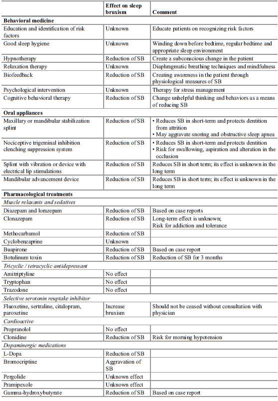
From Contemporary oral medicine 2019 The management of SB aims mainly to reduce the damage and consequences of SB rather than reducing the muscle activity. Behavioural medicine: Most of these concepts are based on expert opinion and lack long term results. Avoidance of triggers such as cigarettes, caffeine, alcohol and illicit drugs can be beneficial. Habit awareness and reversal techniques for AB will reduce attrition of the dentition. Relaxation techniques, good sleep hygiene and hypnotherapy may be useful for SB and AB.  Oral appliance: First line therapy for SB associated with TMD. Mechanism of action is under unresolved debate. Current evidence suggests that oral appliances decrease SB transiently for the first 2 weeks of wear with SB continuing in the long term. Changes to occlusion may occur as a side effect and may not be tolerate by those with an active gag reflex. The use of maxillary stabilisation splints may increase the AHI and risk for snoring due to reduced to...
A post on bruxism (part 3)

From Contemporary oral medicine 2019 Signs of SB: -Tooth wear: Erosion most commonly causes excessive tooth wear with attrition following closely. Attrition can be charaterised by flat planes on teeth i.e wear facets, with planar enamel wear and shallow dentine wear with well defined margins in the enamel of incisal edges or with step like areas on the palatal aspects and equivalent facets on all opposing teeth. In contrast, erosion tends to damage dentine more deeply causing cupped lesions and loss of surface anatomy. Bed partner or parent reports of tooth grinding sounds can also be helpful in diagnosis. -Masseter hypertrophy: This is a benign, asymptomatic enlargement of one or both masseter muscles. There is a slight male predominance with an average age of 30 years. -Tongue indentation (Tongue scalloping): This has been associated with bruxism, more commonly clenching. It has been suggested that Tongue indentations may be caused by macroglossia secondary to systemic amyloid...

A post on bruxism (part 2)

From Contemporary oral medicine 2019 The etiology of SB is largely unknow but current theories hypothesise that SB is centrally mediated probably in the brainstem and has a multifactorial etiology. Sleep arousal is a brief awakening from sleep (3-15s) characterised by increased EEG, autonomic, cardiac and muscular activities without a complete return to consciousness. These normally occur <15 times an hour in response to external or internal stimuli. The association between sleep arousals and RMMA is well estabilished but they shouldn't be considered as the only cause or trigger of SB as they may be the window that allows RMMA during sleep. Additionally, arousals represent the end result of a multitude of physiological events involving the SNS, movement and respiration that may be more to blame. Genetics and familial predisposition: The level of evidence supporting a genetic predisposition to SB is low however there is a high proportion of SB subjects who have a family mem...

A post on bruxism (Part 1)

From Contemporary oral medicine 2019 Bruxism is a repetative jaw muscle activity characterised by clenching or grinding of the teeth and/or bracing or thrusting of the mandible. Sleep (SB) and awake (AB) bruxism are separate in their presentation and pathophysiology however they may overlap in some individuals. AB tends to occur in the form of clenching and bracing or thrusting of the mandible. Grinding can be seen but it is milder than that seen in SB unless the patient has a neurological condition. There seems to be a female predilicition with rations M:F between 2:3 and 1:3. AB is associated with stress, depression, addictions, neurological disorders such as Huntington's disease and congitive impairments such as in Rett's syndrome, Down syndrome and ASD. It is also significantly influenced by lifestyle. Management of AB is initially based of the recognition of awake clenching. This may involve lifestyle changes, habit reversal training, relaxation, hypnosis and biofeedba...
Today I had an interesting occurrence whereby there was a sinus infection associated with a dental infection. While I was going through routine consent, he responded to the risk of OAC by saying he has had recurrent sinus infections in the past. he has apparently seen his GP who found nasal polyps and has recurrent sinus infections that he is prescribed antibiotics for. On a side note it is important not to take everything the patient reports as a fact. One would hope that the GP offered more investigation i.e referral to an ENT rather than symptomatic relief from antibiotics. Who knows, maybe the patient was offered this but declined or maybe he sees a different GP every time. Additionally, on the PA it did seem as though the palatal root was close to the maxillary sinus with a periapical radiolucency involved. These should be warning signs to get prooper consent form the patient and inform them of the possible adverse outcomes of treatment and the possible follow up strategies includ...

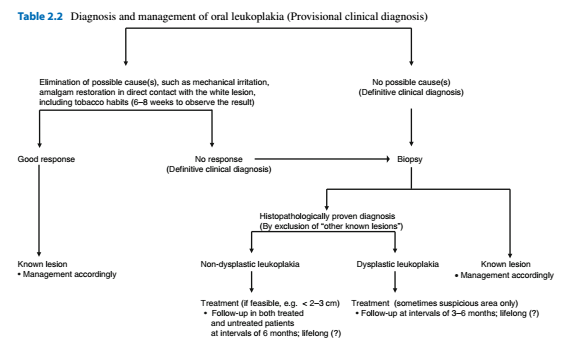
Leukoplakia and other White lesions

Image
Alveolar ridge keratosis White changes of the retromolar region or the alveolar mucosa where teeth have been extracted. Asymptomatic white, homogeneous discolouration of the mucosa Caused by direct irritation from foods against the edentulous ridge by opposing teeth Benign lesion. If believed to be alveolar ridge keratosis then no treatment other than follow up Aspirin burn Superficial burning of the oral mucosa due to the local application of aspirin or paracetamol. White, non wipeable discolouration of the mucobuccal fold, buccal mucosa or border of the tongue Cease drug, Should heal the lesion within a week or two Contact lesion Benign whitish, sometimes erythematous or mixed red and white lesion due to chronic, direct contact with a dental restoration (Usually amalgam) Diagnosis by removal or the amalgam restoration and replacement. Should result in resolution of the lesion within 2-3 months Usually buccal mucosa and borders of the tongue Frictional lesion (Fri...

Cysts of the soft tissues

Epidermoid cyst Rare May manifest during infancy or childhood Most commonly midline of FOM Treatment with enucleation, recurrences are rare Heterotropic gastrointestinal cyst Extremely rare Usually present at birth or soon after Cystic swelling in the anterior floor of mouth Enucleation, rare recurrence Lymphoepithelial cyst (Oral tonsil) Rare Any age  Yellowing, circumscribed swelling, asymptomatic Floor of mouth and ventral tongue Nasolabial cyst Rare Mainly diagnosed during adulthood Paramedian swelling in the upper mucobuccal fold. May show some radiographic erosion. May see swelling of nasolabial groove and bluish swelling intranasally Enucleation through an intraoral approach Mucous retention phenomenon (Mucocele, Ranula) Traumatic obstruction of the duct Common, any age Bluish, non painful, recurre Mainly Lower lip (Mucocele) or floor of mouth (Ranula) involving the sublingual gland Plunging ranula herniates through the mylohyoid muscle pr...

Imaging of the maxillary sinus

There are multiple methods for viewing the maxillary sinus including: 1. Panoramic radiography 2. Water's view 3. CBCT 4. MRI Panoramic radiography has a focal trough that closely resembles the dental arch and so pathologies and abnormalities outside of this focal trough will not be properly imaged. Fluid levels often found in acute sinusitis are not well demonstrated in OPGs. Therefore MRI and CBCT 3D imaging are the gold standard for sinus imaging. We should therefore strive to detect abnormal symptoms and abnormalities on OPG radiographs and provide a diagnosis to avoid unneccessary specialist referrals and diagnostic tests. -Inflammatory diseases Sinus inflammatory conditions are most visible on an OPG when they are on the floor of the maxillary sinus most commonly the mucous retention phenomenon -Mucous retention phenomenon Smooth dome shaped swelling of the mucosa with homogeneous density Rarely symptomatic Requires no treatment Has no relationship to sinus obs...

Always check the soft tissue!

Today I missed an ulcer on the patient's lower lip, on further investigation it turns out the patient has had that same ulcer for a number of years and a previous clinician had recommended a biopsy which she didn't return for. Missing this was a mistake and I need to try harder to check the soft tissues and pay attention to detail. To describe lesions use: 1. Site a. anatomic location (e.g tongue, floor of mouth, palate) b. Adjacent features (E.g adjacent to 34 tooth) c. Unilateral/bilateral d. Localised/Generalised 2. Colour 3. Morphology a. Diameter (mm) b. Single/multiple c. Separate/Coalescing d. Raised/flat/depressed e. Fluid filled/tissue filled/texture f. Margins (Regular/irregular, Raised/smooth, Colour)