Posts

Showing posts from April, 2016

Cervical burnout

Currently trying to find out more information about cervical burnout (CB) and its differentiation from root surface caries (RSC) as I have had trouble distinguishing the two in the past. From what I can gather, -CB appears on the mesial and distal surfaces of teeth in the cervical region - It is a diffuse (ill defined) radiolucent (blacker) area and is bounded by the CEJ and the alveolar ridge -It may be due to overexposure of the film which can "burnout" the thinner sections of teeth. i.e there is lower absorption of the xrays by this anatomical area so if the exposure is high enough, the film in this area will totally "blacken". -It is optically more prominent due to the contrast with the adjacent radioopaque bone and enamel. -RSC has a similar radiolucent bowlshaped area on the radiograph. However, It will generally have a loss of tooth structure i.e in CB there is an intact tooth surface radiographically whereas this is not intact in RSC. Also, CB will sit
Palatine torus/Torus palatinus

From a 1st year UQ dent lecture  -Located close to midline of palate   -Can be either unilateral or bilateral   -Autosomal dominant with virtually 100% penetrance This means that almost everyone that carries this gene will exhibit a torus to some degree. Penetrance refers to the proportion of the population carrying the gene that express its effects. If a particular gene has low penetrance, then even though many people carry it, relatively few will show it in the phenotype. Torus Palatinus also has variable expressivity. This means that, even though most patients who inherit the gene will exhibit some signs of a torus palatinus, the degree to which they express the gene is variable, so there will be a wide range of severity within the population of affected patients.   -It usually becomes apparent around the time of puberty -More commonly seen in females than in males.   -Clinically, it is not considered dangerous. However, if a denture is required, then the torus m

Paedo Exam

It is important to look at the occlusion in every patient but especially in child patients as early diagnosis of occlusal anatomy will allow timely orthodontic intervention. Note: -Overbite: normally 3-4mm or 30% but look for openbites, deep bites, incomplete overbites (where there is overlap but anterior teeth don't contact). Class 2 often has a deep overbite -Overjet: normally 2-2mm. Increase may suggest thumbsucking. OJ may be indicative of angles class. Edge to edge or crossbite in class 3, reduced in class 2 div 1, increased in class 2 div 2. - Angles class -Crossbites: anterior, posterior lingual crossbite or buccal crossbite (scissor bite) -Excessive wear-> interferences -Presence of canine bulge -Missing teeth/Later eruption of permanent teeth/exfoliation of deciduous teeth

Removing edentulous impression trays

Firstly, it is useful to mark the vibrating line using an indelible marker like thomspon sticks so you know if you have over or underextended the tray. this will transfer to the 2nd impression and ensure that the correct extension is made of your wax rim. Suction can be quite strong with these impressions and to ease removal of the tray, push the handle tissue ward to allow the posterior section of the tray to disengage first and this will break the seal.

Sectional matrix bands

Short post here, When restoring and adjacent MO and DO with sectional matrix bands, it is useful to place both matrix bands at the same time. if you pace one at a time then you may find the first restoration may sit somewhat into the 2nd cavity. this may result in a poor concave contour on the 2nd restoration. the first band can be removed after the first restoration as we want to minimise the thickness of the band to get the best contact.

Toothbrush abrasion

It is quite common to see toothbrush abrasion in this day and age. Patients who have nervous disorders or who have had previous caries experience may go overboard because they feel as though they are not cleaning well enough. Some points to raise with them are: -You're brushing too hard. To help --> electric toothbrushes may stop if you press too hard (although i still think they let you press too hard before stopping) and they just have to hold the toothbrush against the tooth while it's working. You don't need a scrubbing action to clean teeth -->Use a soft toothbrush --> slow down. People in a rush when brushing tend to press harder and scrub faster --> avoid whitening or smokers toothpaste. They tend to be more abrasive "Tubes run down the exposed root and cold water and air stimulate these tubes." -Symptoms in normal people should subside with mineral deposition in tubules. In their case, either toothbrush abrasion or acidic diet/reflux

Calcifications in OPGs

Image
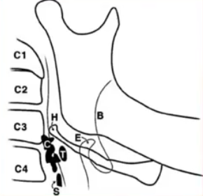
Watched a CPD video on OPG calcifications. The main groups were: -TMJ calcifications -Sinus calcifications -Glandular calcifications -Lymph node calcifications Why do they occur? 1. Dystrophic calcification: Most common in OPGs, Damaged/degenerating tissue. Often a sign of ageing but can be pathologic 2. Metastatic calcification: Rarest: Increased plasma levels dt metabolic disorder e.g hyperparathyroidism 3. Calcinosis: Vascular disease where there is calcification in the sub-, cutaneous or deep ST. Assoc with collagen vascular disease 4. Other Where? 3 broad areas 1. Intracranial: Brain, pituitary 2. Maxillofacial: TMJ, Sinus, Salivary glands, lymph nodes 3. Neck: Lymph nodes, ligaments, vascular Maxillofacial TMJ: -Osteophyte fracture: Osteoarthritic changes, breaks off and causes "joint mites" in the joint space - Ankylosis: Loss in height of condylar neck and change in morphology (loss of outline and hypoattentuating area in the joint space) Rare but

Injection tips

Just watched a CPD video by Dr. Townsend from Uni of Adelaide. Some tips I picked up: -Painful injection technique involves pulling the tissues as taut as possible and having a slow injection technique -topical anaesthetic should be applied minimally (you only need a little bit) and on dry mucosa. An effort should be made to keep saliva from washing the topical off as the taste is poorly handled. - IDB technque:    Level: 1cm above the lower molars, halfway between the upper and lower teeth when the mouth is wide. At the level or just above the level of the coronoid notch which is the most concave portion of the anterior border of the ramus    Point: Just anterior to the Pterygomandibular raphe (Join of superior constrictor muscle and buccinator muscle) which is signified by the Pterygomandibular fold Medial to the buccal pad (not fat but fibrous tissue) Medial to the tendonous portion of the temporalis muscle at the anterior coronoid process. This results in a point in the pt

Bonding of indirect restorations

Indirect restorations are constructed outside the mouth and many steps are added on compared to direct restorations. Therefore, the utmost care is taken to ensure a good fit as poor fit will result in remake and lost time and money. But even with proper care, it is important to follow the correct clinical steps to ensure longevity of the restoration. I had a flat-prep, porcelain emax onlay constructed that wasn't seating (tooth 25-lingual was seating but was rocking on the buccal). Potential causes of this issue that were running through my head: 1. impression distortion, 2. temporary wasn't fully removed (structur with spot etch, bond and flowable resin cement) 3. Incorrect construction of restoration Solution: I had isolated the tooth with single tooth rubber dam. The restoration was contacting the rubber dam (due to its thickness and was preventing full seating. I modified the rubber dam position to stretch to include the 24 and cut a slit on the distal rubber dam to