Posts

Showing posts from May, 2016

Perseverence

-Don't worry about money, Make your dentistry as good as you can and the money will follow -Don't start with speed, build good habits and increase your skills. Rubbish in= rubbish out. -Don't worry about where your peers are at. It's not a competition, it's life. Focus on making yourself as good as you can be. -You have worked hard to get where you are today, don't let patients think they know more than you. In the end their treatment decision lies with themselves but they shouldn't force you to do something you don't believe is right.
Dental trauma-Splinting

Splinting of teeth that have undergone physical trauma allows the periodontal ligament to undergo repair without the trauma of mobile teeth. It is necessary in mobile and displaced teeth i.e Subluxation (in some cases), Lateral luxation, intrusion, extrusion, avulsion, horizontal root fractures, alveolar bone fractures. An easy way to remember splinting times is that hard tissue injuries (Root fracture, lateral luxation (damage to bone), alveolar bone fractures) will require 4 weeks of splinting. An exception is cervical 1/3 root fractures which will require 4 months of splinting. Non-crushing injuries (avulsion, extrusion) are actually less damaging to the periodontal ligament and so will require up to 2 weeks of splinting to allow repair. Intrusion depends on the treatment provided. In very young teeth with minimal intrusion, spontaneous reeruption may occur that will not require splinting. Orthodontinc repositioning and surgical repositioning will require splinting times (4 weeks)

Notes

Stay back at the end of the day and review your notes and xrays. You owe it to the patient and yourself to have good notes and not miss anything diagnostically. Always ask yourself: "if this was taken to court, would it be defensible?" Professionally written notes are important as someone reading back on them can question your level of knowledge etc. Everything needs to be in writing or it didn't happen. If you got correspondence from the patient's GP, then that needs to be in writing in a formal letter from them. But don't get that during their work hours, you can send off an email and they will reply at the end of their day (as you can be disturbing their daily schedule)

Denture review

In a review appt for full dentures (especially immediate dentures- as they have more going on), if a patient presents with a sore spot, Always check the occlusion first before hacking away at the denture. If they have an occlusal interference or CR is incorrect, the patient may be causing a denture base shift in function which will require occlusal adjustment or remake rather than a denture intaglio adjustment.
Be wary of performing irreversible procedures on patient justified just because they think they won't undergo elective procedures in the future. They could win the lottery the next day and suddenly they have no tooth left to work on. Start conservative and don't jump to irreversible treatment unless there is an overriding reason (they're absolutely sick of the condition of a tooth and want it out/ are going away for a long time and will not have access to a dentist. If their dental issue is a minor inconvenience e.g extra cleaning for food packing, then they can live with it until they are ready to extract or can afford more pricier treatment e.g ortho. Extraction of a lateral instead of a palatally placed canine to stop food packing is preferrable as the canine is harder to extract and will last for life and the canine can be odontoplastied to blend in as a lateral.

Post endo molar pain

A root canalled upper molar exhibiting pain post treatment may be due to a missed MB2 canal. This requires reaccess and debridement of the missed canal. this may need a referral to the endodontist.
When looking at out of place dental radiolucencies, check for enamel abnormalities e.g enamel fractures that may be on the strangest places and can appear to be carious on the x ray. Radiolucent areas under restorations may be voids or bases. Track cracks with your probe. Separation indicates a need for intervention and if they track subgingivally on the most posterior tooth this requires referral to an endodontist to assess the tooth for suitability for restoration.

Clean working area

Cleanliness is important in a work space. Patients will see disorganised work benches and wonder if your work in their mouth will be disorganised. Ensure you and your D.A cleans unneeded scraps from your bracket table and ensure blood soaked items are out of sight e.g in a cup to avoid the patient seeing them. The area on the floor is important as well so ensure that it is regularly cleaned.

Advice from Dr. Kelsey

Follow your dreams; fulfill your destiny. It won't happen on its own.  What a legend.

Tips from 3rd may 2016

Started with Dr Kelsey today and have had a good experience so far. Here's what I picked up: -To test if a restoration is high, you can apply a localised bite force with a thick bit of cotton pellet and place it on the resto surface with some tweezers. On adjacent MO and Do restorations, you can place it on the interproximal area and then narrow it down by placing it on individual restorations - To find a cracked cusp, you can give the patient a frac finder and tell them to chew around with it till they find the sore cusp. then leave it there for you to take a look. -Pain on release with a cracked cusp occurs as biting causes separation allowing fluid to enter the cracked space. Release of pressure will cause rebound of the tooth and will force fluid down the dentine tubules which are quite raw as they are normally unexposed. - An explanation--> "There is enamel covering the tooth which is non sensitive with no nerve endings and underneath is the dentine which is sensit...

Real image, double real image, ghost image

Image
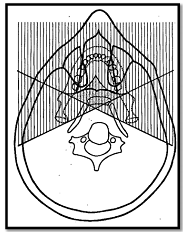
OPG beams originate from behind the patient. As it is 3D projected onto 2D there are many superimpositions. These may be real images, double real images, ghost images, soft tissue, airway spaces. Double real images are formed in the central diamond area as the beam will pass through here twice. -One image is the mirror image of the other -Both images are real -Each image has similar proportions -Each image has the same location on the opposite side -Only occurs with midline objects e.g Hard and soft palate, palatal tori, body of the hyoid, epiglottis, cervical spine Ghost images are formed when the object is between the xray source and the centre of rotation (behind the centre of rotation i.e white area) -Has the same general shape -Appears on the opposite side of the radiograph and is a mirror image -Appears higher up as the xray beam is angled slightly upwards -Appears more blurred -Vertical component is more blurred than horizontal component -Vertical component is...

Radiographic interpretation

Passing through an old radiograph lecture and wanted to list a few hints: -PDL space is thinnest near the middle of the root and wider near the alveolar crest and apex (As fulcrum is where the PDL is thinnest) -Lamina durat is the thin radioopaque area representing the alveolus wall (Socket wall where sharpeys fibres of the PDL insert into bone). This is more dense than surrounding trabecular bone and will be thicker and more prominent with increasing occlusal stress (as a compensating mechanism) -The crest of the alveolar bone is considered normal height if it sits within 1.5mm of the CEJ

Radiographic appearance of tumours

Odontogenic tumours can be classified as benign or malignant. Benign tumours: -Well defined but may not be corticated (margin discernible but border may not be more radioopaque than adjacent bone) -Slow growing therefore: -Displaces teeth -Causes blunt root resorption -Displaces IAC -Expansion and thinning of mandibular cortex Malignant tumours: -Aggressive and fast growing -Ragged or Pencil sharpening root resorption (Mottling of roots) -Erodes cortex of IAC (as opposed to deflection) -Expansion and ragged erosion of adjacent cortex -Loss of lamina dura-> widening of periodontal ligament space -Periosteal reaction