Real image, double real image, ghost image
OPG beams originate from behind the patient. As it is 3D projected onto 2D there are many superimpositions. These may be real images, double real images, ghost images, soft tissue, airway spaces.
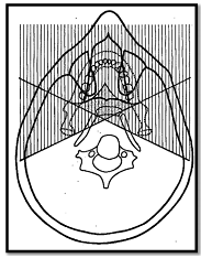
-One image is the mirror image of the other
-Both images are real
-Each image has similar proportions
-Each image has the same location on the opposite side
-Only occurs with midline objects e.g Hard and soft palate, palatal tori, body of the hyoid, epiglottis, cervical spine
Ghost images are formed when the object is between the xray source and the centre of rotation (behind the centre of rotation i.e white area)
-Has the same general shape
-Appears on the opposite side of the radiograph and is a mirror image
-Appears higher up as the xray beam is angled slightly upwards
-Appears more blurred
-Vertical component is more blurred than horizontal component
-Vertical component is much more magnified, horizontal dimensions may be less severely magnified. It is larger as the beam diverges and the ghost image is encountered behind the centre of rotation.
-Main Ghost image is the cervical spine and angle and ramus of the mandible
Comments
Post a Comment
Please leave a comment and let me know what you think or if there are any topics you would like covered in the future