Posts

Showing posts from January, 2017

Extraction positioning

Image
Patient position Maxillary extractions: Mouth must be the same height as the dentist's shoulder. Angle of the dental chair back and the floor should be around 120 degrees. The occlusal surface of the maxillary teeth should be 45 degrees to the horizontal when the patient's mouth is open. Mandibular extractions: The chair is positioned more upright so the angle of the chair back is 110 degrees. The occlusal surface of the mandible must be parallel to the horizontal when the patient's mouth is open. Dentist positioning Right handed: To the front and right of the patient. For anterior and mandibular teeth the dentist should be in front of the patient or behind them and to the right Left handed: front and left of the patient. For anterior and mandibular teeth the dentist should be behind the patient and to their left.
Boxing impressions to pour up

Boxing impressions can be beneficial before pouring up casts because upon removing the impression from the cast the cast will be of ideal shape and will require minimal adjustment. Inverting impressions onto a stone base can be harmful as if the stone base is too firm it can distort the stone overlying the impression especially over unsupported areas. Also, water in the stone mix rises upwards against gravity so inverting the impression will result in a higher concentration of water at the working surface resulting in a weaker cast predisposed to chipping. Boxing allows for the impression to be poured without inverting. Steps: 1. If it is a lower impression, the lingual area must be blocked out with a sheet of red wax. Trim a wax sheet to the approximated shape of the lingual area and seal 2-3mm from the sulcus depth with sticky wax or a hot wax knife. 2. Soften a sheet of red wax and fold over the long edge once. Cut a thin strip ~2mm wide. This will result in a strip ~3mm thick ...

Managing endodontic cases of differing complexity

General guideline of RCT: 1. Analysis of the specific anatomy of the case 2. Canal scouting 3. Coronal modifications 4. Negotiation to patency 5. Determination of working length 6. Glide path preparation 7. Root canal shaping to desired size 8. Gauging the foramen, apical adjustment   Pay attention to the following steps with these difficulties Cases with relatively low complexity: Step 1: Analysis of the specific anatomy of the case Step 4: Negotiation to patency Step 5: Determination of working length Simple cases probably have larger pretreatment canals. Consider using larger files for working length determination with an apex locater. The better the adaptation of the tip of the instrument to the foramen, the more accurate the length determination. Step 6: Glide path preparation Step 7: Root canal shaping to desired size The more recently advocated simplification of using a single-file, single-length technique may work well in this anatomy, where a single rotary or recip...

Canal negotiation to working length

Precurving the negotiating  instrument at canal negotiation is not essential but may be needed in cases of moderate to  severe curvature ; in case the file stops before the estimated working  length, either the file should be precurved to be able to bypass an  impediment or more space needs to be created midroot or in the apical third. These two conditions are defined by the tactile feedback the clinician gets from the file; a loose resistance indicates the presence of a ledge or acute curve, while so-called rubbery resistance suggests the presence of a tight canal space or the presence of compacted soft tissue that prevents the file from penetrating deeper into the canal.    West  differentiates four specific conditions that require technique modifications: 1. Apical blockage 2. Mismatch between canal curve and instrument 3. Too large file tip 4. Presence of restrictive coronal dentin All these conditions may be remedied by either changing...

Leukoplakia and other White lesions

Image
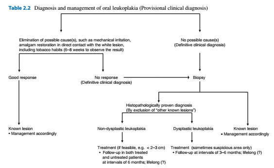
Alveolar ridge keratosis White changes of the retromolar region or the alveolar mucosa where teeth have been extracted. Asymptomatic white, homogeneous discolouration of the mucosa Caused by direct irritation from foods against the edentulous ridge by opposing teeth Benign lesion. If believed to be alveolar ridge keratosis then no treatment other than follow up Aspirin burn Superficial burning of the oral mucosa due to the local application of aspirin or paracetamol. White, non wipeable discolouration of the mucobuccal fold, buccal mucosa or border of the tongue Cease drug, Should heal the lesion within a week or two Contact lesion Benign whitish, sometimes erythematous or mixed red and white lesion due to chronic, direct contact with a dental restoration (Usually amalgam) Diagnosis by removal or the amalgam restoration and replacement. Should result in resolution of the lesion within 2-3 months Usually buccal mucosa and borders of the tongue Frictional lesion (Fri...

Cysts of the soft tissues

Epidermoid cyst Rare May manifest during infancy or childhood Most commonly midline of FOM Treatment with enucleation, recurrences are rare Heterotropic gastrointestinal cyst Extremely rare Usually present at birth or soon after Cystic swelling in the anterior floor of mouth Enucleation, rare recurrence Lymphoepithelial cyst (Oral tonsil) Rare Any age  Yellowing, circumscribed swelling, asymptomatic Floor of mouth and ventral tongue Nasolabial cyst Rare Mainly diagnosed during adulthood Paramedian swelling in the upper mucobuccal fold. May show some radiographic erosion. May see swelling of nasolabial groove and bluish swelling intranasally Enucleation through an intraoral approach Mucous retention phenomenon (Mucocele, Ranula) Traumatic obstruction of the duct Common, any age Bluish, non painful, recurre Mainly Lower lip (Mucocele) or floor of mouth (Ranula) involving the sublingual gland Plunging ranula herniates through the mylohyoid muscle pr...

Analysing the aetiology of tooth wear

Image
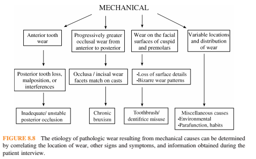
Mechanical wear: Restorations tend to wear at the same rate as surrounding tooth structure. Displays sharply defined peripheries that can be matched on articulated diagnostic casts. Frequently asymptomatic and patients may report parafunctional habits Anterior wear greater than posterior wear: Posterior tooth loss, malposition or interferences Inadequate or unstable posterior contacts-> anterior teeth taking more force-> anterior wear and loss of vertical dimension Posterior occlusal premature contacts-> anterior posturing-> increased function-> wear Progressively greater wear on the anterior teeth: (Exception in anterior open bite). Bruxism. May also show grooving of the lateral borders of the tongue, evidence of cheek biting, fracture of porcelain restorations. Cupping or cratering of the occlusal surfaces can occur once the enamel has been perforated. Facial surface of canines and premolars: Excessive toothbrushing resulting in a sandblasted appearnce with a...

Imaging of the maxillary sinus

There are multiple methods for viewing the maxillary sinus including: 1. Panoramic radiography 2. Water's view 3. CBCT 4. MRI Panoramic radiography has a focal trough that closely resembles the dental arch and so pathologies and abnormalities outside of this focal trough will not be properly imaged. Fluid levels often found in acute sinusitis are not well demonstrated in OPGs. Therefore MRI and CBCT 3D imaging are the gold standard for sinus imaging. We should therefore strive to detect abnormal symptoms and abnormalities on OPG radiographs and provide a diagnosis to avoid unneccessary specialist referrals and diagnostic tests. -Inflammatory diseases Sinus inflammatory conditions are most visible on an OPG when they are on the floor of the maxillary sinus most commonly the mucous retention phenomenon -Mucous retention phenomenon Smooth dome shaped swelling of the mucosa with homogeneous density Rarely symptomatic Requires no treatment Has no relationship to sinus obs...