Posts

Showing posts from 2017

Restoration of endodontically treated teeth

Typically, root canal treatment is initiated because of deep caries or trauma, both of which often result in extensive loss of tooth structure. Additional tooth tissue is removed for endodontic access, cleaning and shaping of the root canal, and post space preparation, further reducing the structural integrity of the tooth and decreasing its resistance to fracture.   At one time it was believed that endodontically treated teeth are inherently more brittle and susceptible to fracture.   Subsequent research has shown that the dentin of endodontically treated teeth exhibits mechanical properties equivalent to that of untreated teeth. It has also been proposed that a portion of the sensory feedback mechanism is lost when the neurovascular tissue has been removed from the tooth in the course of endodontic therapy. Clinically, this means that the patient can inadvertently bite with more force on an endodontically treated tooth than on a vital tooth because of the impaired ...

Cracks, cracks, cracks

Image
Today I had a case that taught me not to focus on the obvious cause of pain. A patient came in complaining of pain on the 25. the 24 had previously been filled by another dentist and I assume it must have been a carious exposure. Checking the xrays before the patient arrived I settled on the 24 as the probably cause of pain. She presented and was very sure that it was the 25 painful to hot and cold. There was an MOD amalgam in the 25 that appeared sound. I still thought it was the 24... Cold test was very painful for both but more so for the 25. The curiosity was that there was an exaggerated response to pain on the 25 as it appeared sound. I was very ready to extract the 24, so much so that the needle was right near the tooth for LA. I decided to test the teeth once more and again a more exaggerated response from the 25. Still unsure if the patient was confused, I did a heat test for the first time. Rubber dam isolation of the 25 and washing of the tooth with warm water from a monoje...

Ankylosed and submerged primary molar

In the absence of ankylosis, primary molars without permanent successors may function for many years before exfoliation, preserving alveolar bone height and width . If a periapical radiograph shows flat bone levels between the submerged primary molar and adjacent teeth, the tooth may be maintained, preserving alveolar bone until facial growth is complete, and an implant can be placed. In this case, the mesial and distal surfaces of the mandibular primary molar can be disked to achieve premolar width. The mesiodistal width at the cementoenamel junction measured on a bitewing or periapical radiograph provides a good guideline for the amount of reduction, as does comparison to the contralateral side. The average width of the mandibular second premolar is  7.5 mm. Seven millimeters has also been recommended as the width to attain. This size can be marked with a pencil or marking pen on the occlusal of the primary molar to provide a guide for reduction. After administration of local ane...

Local Anesthetic Considerations When Treating Patients with Oral Infections

Products of inflammation lower the surrounding tissue pH (e.g., purulent exudates have a pH of 5.5-5.6). At this more acidic pH, the numbers of base molecules necessary for passage of the anesthetic into the nerve membrane may be significantly reduced. Inflammatory exudates may also enhance nerve conduction action potentials, making blockage of sensory nerve impulses more difficult. In addition, blood vessels in the area of inflammation may be dilated, leading to a more rapid uptake of the anesthetic agent from the area of injection. These changes can lead to delays in onsets of anesthesia, inadequate depths of anesthesia, and the potential for local anesthetic blood levels to be elevated.  Needle tract  infection  is a potential complication of injection into  infected  tissues. Although penetration into  infected  tissues can be avoided by using more distant regional nerve blocks, whenever there is a possibility of a needle having passed through...

Curing light ADA video

-Roast chicken analogy: Put a chicken in the oven at the highest heat and when it looks good on the outside is it cooked well? -Irradiance: power/area -Power->Watts -Bulk filling: Light tip has to cover the restoration completely. Some MFRS are smaller diameter which is smaller than a molar M-D distance. -Uncured CR, Uncured monomers at the deepest part esp pulpal area-> Doesn't have the physical properties that MFR intends.  "Pt is biting down on a sponge" common reason for Cr to fail. Low conversion rate causes leakage of monomers and photoinitiators. -Overcuring can cause damage: cook the pulp, damage the gingiva, Follow MFR instructions. Can cure longer but should have a break e.g 5 seconds or blow air over tooth -Ramp cure and pulse lights, no beneficial effect shown. Ramp effect needs to increase over minutes not seconds to be actually effective in reducing problems -Variation within composites: Shade-> can have maximum increment size differences -C...

Mental blocks

Today I had to extract lower 3-3 for the insertion of a full lower immediate denture. I was hesitant to give bilateral IDBs and asking around it seemed as though the other dentists would be as well. I got around it by giving bilateral mental blocks and lingual infiltrations. There were no premolars for intraoral landmarks so I palpated extraorally then intraorally to find the mental foramen. I retracted the soft tissues with my finger to have an intraoral reference to penetrate the mucosa. Lingual infiltrations I placed around the lateral incisor area and it seemed to be sufficient to anaesthetise one tooth either side. The OPG also helped in approximating the position distal to the canines.

Composite resin protocol

This is the composite resin protocol I've been using recently including instruments: 1. LA, Rubber dam, prewedge and cavity prep (Lately I've had difficulty with larger spaces between teeth, the largest wooden wedge hasn't binded and stayed in place. I may try a plastic wedge in the future. It has the benefit of sitting down into the gingival sulcus and inverting the dam) 2. Matrix band and burnish against the next tooth a) 1-2 walls missing interproximally: Sectional matrix band/s. If the buccal and lingual extensions are too severe and the use of a ring will crush the band then use a wooden wedge to seal the gingival margin. The band can be adapted into the gingival sulcus for adaptation against the tooth but most likely i'll need to trim the excess material post placement b) 3 walls missing: Tofflemire band and wooden wedges or plastic wedge and separating ring c) 3.5-4 walls missing: Automatrix and wooden wedges 3. Bonding procedures 4. With the composite re...

Don't let patients dictate your treatment.

I recently had a patient that required extraction of a heavily filled lower 6. The last xray we had was a PA 2-3 years old. I told him that I would like to take a new Xray to plan the extraction. HE kicked up a big fuss about the cost of the xray. I compromised and said I would take the tooth out with the old xray. Big mistake. The crown fractured and the roots kept fracturing with elevation. The extraction would have been just as difficult with the updated xray but medicolegally I don't think I would have a good defence if things went sour. I took a PA halfway through anyway to check the progress of the extraction. It scared me because at the angle I took it, the IAN overlaid the roots. Then I realised I should have taken the xray at the start. In the future if a patient kicks up a fuss about the cost of necessary diagnostic tests I would either straight up refuse to perform the treatment or give the xray for free. It's 30 seconds work to save a lot of headache down the track....

Photography

Image
-Full smile For this shot, a point-and-shoot camera should be switched to macro mode; DSLR macro lenses should be used at approximately 1:2 magnification. Attempt to take the photo from directly in front of the patient, avoiding a downward or upward angle of view. The patient should exhibit a natural smile, with framing of the photo extending from the right to left corner of the mouth. Point of focus for this shot is on the central or lateral incisors. The horizontal midline should be the incisal plane; the vertical midline should be the anatomic midline.  -Anterior retracted Patient seated with dentist in front. Cheek retractors used with the cheeks pulled outwards away from the teeth. A common mistake is to pull the lips outwards and backwards which pulls the buccal area inwards. Use the largest retractor possible to avoid the centre of the upper and lower lip from appearing in the photo. Air dry the teeth. the teeth should be in MIP but they can be slightly apart to aid in...

Code 627

Preliminary restoration for crown is the code 627. I believe that this code is too all encompassing. I restored a tooth with composite resin today, lower molar 5 surface. If my core buildup was only 3 or 4 surface the cost would be the same, I think that there should be an increase in cost for larger buildups which perhaps relates to the cost of the correstponding 5-- code. e.g Half the cost of the 5-- code. Such problems would be less obvious if we used a dual cure composite core buildup system which involves a single layer which would be a timesaver. If I have to buildup the whole tooth in CR with incremements then running costs increase significantly.

Moisture control

Today I placed a large 12MIDL composite resin without rubber dam. Not sure about my reasoning about the lack of rubber dam but it would have been difficult to place due to the subgingival nature of the prep. This is not an excuse however and the patient's inflamed gingiva kept bleeding onto the composite resin. Long term, this increases the risk of staining and secondary caries. I placed a retraction cord but didn't place it interproximally. Instead i placed it flowing onto the gingival margin of the adjacent teeth. I think that the lack of cord in the interproximal allowed bleeding onto the composite resin. If I could restore this tooth again, I would pack cord or teflon tape then rubber dam from premolar to premolar and use floss ties to invert the dam into the sulcus +/- an anterior cord. Also, the contralateral lateral incisor was missing which made it difficult to form anatomy till symmetry. Additionally the ipsilateral canine was buccally and labially positioned. A cont...

Radix entermolaris and Radix paramolaris

Extra root present on lower molars. Non concentric position and easily missed in RCT RE is present distolingually in lower molars but can be located as far messially as the central portion between the mesial and distal roots. RP is present mesiobuccally but can be found centrally between the mesial and distal roots RE Rare in Caucasian populations Max 3-4% RE Eurasian and Indian <5% RE Mongoloid population (Chinese, eskimo and american indians) 5% - >30% RE can be found on and lower molar but occurs most commonly on the 6 and least commonly on the 7. 50-67% bilateral Morphology can vary from a short conical extension to a mature root with normal length and root canal. In general, the RE is smaller than the DB and mesial roots and can be separate from or partially fused RP is very rare. 2% 3rd molar, 0.5% 2nd molar, 0% first molar but other studies have found RP in first molars.  An additional cusp (Tuberculum paramole) may be present on the buccal side in a tooth wi...

ADA patient information sheet series- Cracked tooth syndrome

I've picked up some patient information sheets from work, just going to spend a few posts summarising the key points of these. -Usually occurs on a molar or premolar (in order of likelihood): 1. Upper premolar 2. Lower molar 3. Upper molar 4. Lower premolar Symptoms: -Sharp and erratic pain on chewing or after release of biting pressure. But not all cracks cause pain  -Pain or discomfort when exposed to cold or hot liquids or food -Sensitivity to sweet -Difficulty in pinpointing location of pain -If the crack extends blow the gum, a periodontal pocket may be present -Often a history of other cracked teeth Causes: -More likely in recent times as teeth are retained for longer with larger restorative cycles and teeth more prone to fracture -Stress leading to grinding of teeth especially at night -Time, wear and tear from chewing, grinding and clenching -Chewing on hard foods e.g ice, sweets or pencils -Trauma especially if upper and lower teeth have been rammed to...

Extraction positioning

Image
Patient position Maxillary extractions: Mouth must be the same height as the dentist's shoulder. Angle of the dental chair back and the floor should be around 120 degrees. The occlusal surface of the maxillary teeth should be 45 degrees to the horizontal when the patient's mouth is open. Mandibular extractions: The chair is positioned more upright so the angle of the chair back is 110 degrees. The occlusal surface of the mandible must be parallel to the horizontal when the patient's mouth is open. Dentist positioning Right handed: To the front and right of the patient. For anterior and mandibular teeth the dentist should be in front of the patient or behind them and to the right Left handed: front and left of the patient. For anterior and mandibular teeth the dentist should be behind the patient and to their left.

Boxing impressions to pour up

Boxing impressions can be beneficial before pouring up casts because upon removing the impression from the cast the cast will be of ideal shape and will require minimal adjustment. Inverting impressions onto a stone base can be harmful as if the stone base is too firm it can distort the stone overlying the impression especially over unsupported areas. Also, water in the stone mix rises upwards against gravity so inverting the impression will result in a higher concentration of water at the working surface resulting in a weaker cast predisposed to chipping. Boxing allows for the impression to be poured without inverting. Steps: 1. If it is a lower impression, the lingual area must be blocked out with a sheet of red wax. Trim a wax sheet to the approximated shape of the lingual area and seal 2-3mm from the sulcus depth with sticky wax or a hot wax knife. 2. Soften a sheet of red wax and fold over the long edge once. Cut a thin strip ~2mm wide. This will result in a strip ~3mm thick ...

Managing endodontic cases of differing complexity

General guideline of RCT: 1. Analysis of the specific anatomy of the case 2. Canal scouting 3. Coronal modifications 4. Negotiation to patency 5. Determination of working length 6. Glide path preparation 7. Root canal shaping to desired size 8. Gauging the foramen, apical adjustment   Pay attention to the following steps with these difficulties Cases with relatively low complexity: Step 1: Analysis of the specific anatomy of the case Step 4: Negotiation to patency Step 5: Determination of working length Simple cases probably have larger pretreatment canals. Consider using larger files for working length determination with an apex locater. The better the adaptation of the tip of the instrument to the foramen, the more accurate the length determination. Step 6: Glide path preparation Step 7: Root canal shaping to desired size The more recently advocated simplification of using a single-file, single-length technique may work well in this anatomy, where a single rotary or recip...

Canal negotiation to working length

Precurving the negotiating  instrument at canal negotiation is not essential but may be needed in cases of moderate to  severe curvature ; in case the file stops before the estimated working  length, either the file should be precurved to be able to bypass an  impediment or more space needs to be created midroot or in the apical third. These two conditions are defined by the tactile feedback the clinician gets from the file; a loose resistance indicates the presence of a ledge or acute curve, while so-called rubbery resistance suggests the presence of a tight canal space or the presence of compacted soft tissue that prevents the file from penetrating deeper into the canal.    West  differentiates four specific conditions that require technique modifications: 1. Apical blockage 2. Mismatch between canal curve and instrument 3. Too large file tip 4. Presence of restrictive coronal dentin All these conditions may be remedied by either changing...

Leukoplakia and other White lesions

Image
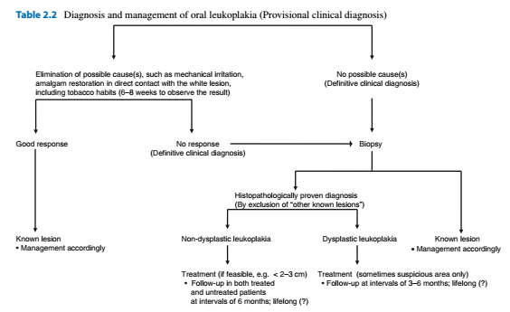
Alveolar ridge keratosis White changes of the retromolar region or the alveolar mucosa where teeth have been extracted. Asymptomatic white, homogeneous discolouration of the mucosa Caused by direct irritation from foods against the edentulous ridge by opposing teeth Benign lesion. If believed to be alveolar ridge keratosis then no treatment other than follow up Aspirin burn Superficial burning of the oral mucosa due to the local application of aspirin or paracetamol. White, non wipeable discolouration of the mucobuccal fold, buccal mucosa or border of the tongue Cease drug, Should heal the lesion within a week or two Contact lesion Benign whitish, sometimes erythematous or mixed red and white lesion due to chronic, direct contact with a dental restoration (Usually amalgam) Diagnosis by removal or the amalgam restoration and replacement. Should result in resolution of the lesion within 2-3 months Usually buccal mucosa and borders of the tongue Frictional lesion (Fri...

Cysts of the soft tissues

Epidermoid cyst Rare May manifest during infancy or childhood Most commonly midline of FOM Treatment with enucleation, recurrences are rare Heterotropic gastrointestinal cyst Extremely rare Usually present at birth or soon after Cystic swelling in the anterior floor of mouth Enucleation, rare recurrence Lymphoepithelial cyst (Oral tonsil) Rare Any age  Yellowing, circumscribed swelling, asymptomatic Floor of mouth and ventral tongue Nasolabial cyst Rare Mainly diagnosed during adulthood Paramedian swelling in the upper mucobuccal fold. May show some radiographic erosion. May see swelling of nasolabial groove and bluish swelling intranasally Enucleation through an intraoral approach Mucous retention phenomenon (Mucocele, Ranula) Traumatic obstruction of the duct Common, any age Bluish, non painful, recurre Mainly Lower lip (Mucocele) or floor of mouth (Ranula) involving the sublingual gland Plunging ranula herniates through the mylohyoid muscle pr...

Analysing the aetiology of tooth wear

Image
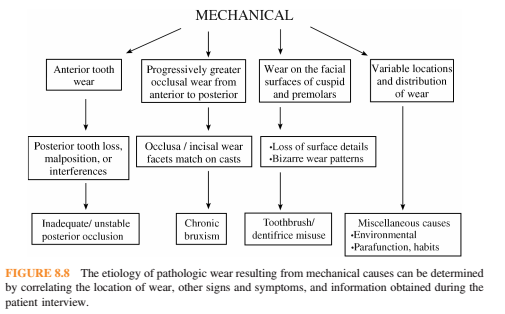
Mechanical wear: Restorations tend to wear at the same rate as surrounding tooth structure. Displays sharply defined peripheries that can be matched on articulated diagnostic casts. Frequently asymptomatic and patients may report parafunctional habits Anterior wear greater than posterior wear: Posterior tooth loss, malposition or interferences Inadequate or unstable posterior contacts-> anterior teeth taking more force-> anterior wear and loss of vertical dimension Posterior occlusal premature contacts-> anterior posturing-> increased function-> wear Progressively greater wear on the anterior teeth: (Exception in anterior open bite). Bruxism. May also show grooving of the lateral borders of the tongue, evidence of cheek biting, fracture of porcelain restorations. Cupping or cratering of the occlusal surfaces can occur once the enamel has been perforated. Facial surface of canines and premolars: Excessive toothbrushing resulting in a sandblasted appearnce with a...

Imaging of the maxillary sinus

There are multiple methods for viewing the maxillary sinus including: 1. Panoramic radiography 2. Water's view 3. CBCT 4. MRI Panoramic radiography has a focal trough that closely resembles the dental arch and so pathologies and abnormalities outside of this focal trough will not be properly imaged. Fluid levels often found in acute sinusitis are not well demonstrated in OPGs. Therefore MRI and CBCT 3D imaging are the gold standard for sinus imaging. We should therefore strive to detect abnormal symptoms and abnormalities on OPG radiographs and provide a diagnosis to avoid unneccessary specialist referrals and diagnostic tests. -Inflammatory diseases Sinus inflammatory conditions are most visible on an OPG when they are on the floor of the maxillary sinus most commonly the mucous retention phenomenon -Mucous retention phenomenon Smooth dome shaped swelling of the mucosa with homogeneous density Rarely symptomatic Requires no treatment Has no relationship to sinus obs...