Posts

Showing posts from December, 2018

Dental sleep medicine series 1: Normal sleep and sleep staging

Image
When treating sleep in a dental setting, it is useful to have a basic understanding of sleep and sleep staging to be able to interpret the results of a sleep study correctly and to be able to communicate effectively with medical colleagues. From Sleep Medicine 6th edition What is sleep Sleep is a reversible behavioral state of perceptual disengagement from and unresponsiveness to the environment. It is also true that sleep is a complex amalgam of physiologic and behavioral processes. Sleep is often associated with behavioural traits such as supine position, quiescence, closed eyes. Parasomnias can occur including sleep walking, sleep talking and tooth grinding. What are the stages of sleep? Two very separate stages of sleep have been identified: REM and Non REM sleep. NREM sleep is separated into 4 substages as defined by the EEG measurement axis (brain waves). NREM EEG is described as synchronous and have characteristics such as sleep spindles, K complexes, and high voltage sl...

Thoughts on the RACDS primary exam

I've applied for subscription to the Royal Australiasian college of dental surgeons. This is the first step towards taking the Primary examinations which is a test of Anatomy, Histology, Physiology, Cell Biology & Biochemistry, Pathology and Microbiology. It is a qualification that displays one's dedication to learning in the field of dentistry and is a prerequisite for many post graduate courses such as those in universities for specialisation and the grad dip conscious sedation that allows you to perform IV sedation in general practice. I am still deciding whether or not to take the exams this year as they are quite full on. My thoughts are: -This is a test of my dedication and willingness to learn. If I can't follow through with the primary exams then I am unlikely to be able to follow through in specialising. -It is better to do this now as I haven't settled down to a long term job yet. -It is better to do this early while I'm not too far away from my u...

Restoring conservative class II cavities

Image
Caries that has just progressed past the DEJ can be a challenge to treat. The milder extent of the cavity preparation can be more difficult to manage than large preparations due to the smaller access and higher risk of voids and maladaptation. After preparation, the cavity prep can often appear like in Figure 1. Class 2 cavity preparation. In shallow caries just into dentine the preparation may be similar to the blue shape. Removal of the orange section may improve visibility and access This small prep although conservative and in the patient's best interest it may cause a sub standard restoration to be placed. The small access affects placement of etch, prime and bond as well as the placement of restorative material and access for finishing and polishing. During preparation, the contact may not be broken which will make the placement of a matrix band difficult. You can get around this by using a stiffer band like a Tofflemire and forcing it through the contact or by widen...

A tip for perio splinting of teeth

Recently I performed a splinting procedure for mobile lower incisors in a perio patient. She presented with significant calculus buildup and radiography showed severe bone loss. Therefore I knew that once I removed the calculus which was bridging the teeth together there would be at least grade 2 mobility. Everstick perio was the product I used. This is because I wasn't prosthodontically adding a tooth onto the bridge and everstik perio has half the amount of fibres as everstick C&B (2000 as compared to 4000) therefore would be thinner and more comfortable. I knew that once I cleaned the calculus off the teeth would be hard to handle as they would be mobile. If done under rubber dam I would not be able to check the occlusion until the teeth were bonded and the dam was off. This may be disasterous as the bridge will be hard to remove if the occlusion is wrong. What I did was ensure the occlusion was comfortable at rest (sometimes it may not be due to traumatic occlusion) The...

Basics of clinical photography notes

Image
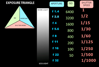
Notes taken from the ADA webinar: Basics of clinical photography by Dr. Peter Sheridan Clinical photographs are used for records and documentation but also professional communication, education marketing etc Issues with intraoral cameras are they have a limited range of views. It is easy to use but poor quality, colour and distortion. Can't integrate the image into the whole mouth context or comparison with other teeth Criteria when choosing cameras -Quality of images -Lightweight -Value -Integrated (can buy all hardware from the same manufacturer) -Ease of use -Depth of field -Special features: eg in built post production software -Longevity Lens should be 60mm not 105mm macro lens. These days, DSLRs are using apsc or dx sensors which are smaller which changes the field of view of the lens. The 60mm lens working at 1.5 crop factor and acts as a 90mm lens and the 105mm lens works as a 150mm lens. The 105mm lens will be heavier, have no depth of field, and will go hu...

Mental nerve blocks

A few thoughts about the technique behind mental nerve blocks: - The path of the IDN through the mandible starts at the mandibular foramen. This is the ideal site for the deposition of anaesthetic for an IDB. as the nerve approaches from superiorly and posteriorly a higher chance of success is gained from injection most posterior and superior. However, too posterior may result in affecting the facial nerve which lies just posterior to the ramus of the mandible. Too far anteriorly will stop the spread on anaesthesia due to a ligamentous attachment to the lingula (sphenomandibular ligament) - Upon entering the mandible, the IDB tracks anterior through the bones near the apices of the lower teeth. At the molar region (if I recall correctly), it sits more to the lingual cortical plate and it shifts to a more buccal position at around the premolar region. This and the denser quality of the bone is why infiltrations have a poorer success rate on lower posterior teeth. At the site of the me...

Case report- Cracked tooth

Image
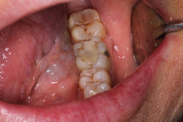
The following is a case report of a procedure where in hindsight I would do things a little differently This patient is a 50 Year old female who presented with the occasional pain to bite down on a lower left tooth. Immediately when I hear this I have an idea in my head as to the possible causes. Firstly the location seems to be fairly reproducible and it only appears to happen when she bites on a certain tooth. This almost completely rules out myofascial pain which would be more constant and deep and would be sore if she bit on any tooth on the affected side. Palpation of the masseter and lateral pterygoid will assist in ruling this out. Do be careful as there is often a secondary myofascial pain from the toothache or from bruxers who often have cracked teeth as a finding. Acute periapical pathology is an option but this would often lead to extended pain after biting and clinical examination should reveal tenderness to percussion which is reproducible. Pulp testing assists in determ...

Some thoughts regarding extraction mechanics and surgical planning

Image
Molar extractions are complex things there's no doubt about it. What makes them so complex is their variation in anatomy ranging from root shape and length as well as curvature, number of roots and the increased surface area of PDL. The surrounding structures add their own complexity with variations in the density of cortical bone as well as surrounding structures of the maxillary sinus and IDN. Therefore it is prudent to plan out extractions properly and have a good idea of how the surgery will go. Here are a few tips I've picked up on surgical planning: -Mandibular posterior cortical bone is dense, don't expect much expansion of the socket -Don't be afraid to raise a flap or go surgical or sectional if it will be easier. Destruction of bone is less of an issue if they aren't planning for an implant. However in the posterior region where extractions are generally more difficult, the volume of bone and soft tissue is less of an issue. Once you remove the first ...