The following is a case report of a procedure where in hindsight I would do things a little differently
This patient is a 50 Year old female who presented with the occasional pain to bite down on a lower left tooth. Immediately when I hear this I have an idea in my head as to the possible causes. Firstly the location seems to be fairly reproducible and it only appears to happen when she bites on a certain tooth. This almost completely rules out myofascial pain which would be more constant and deep and would be sore if she bit on any tooth on the affected side. Palpation of the masseter and lateral pterygoid will assist in ruling this out. Do be careful as there is often a secondary myofascial pain from the toothache or from bruxers who often have cracked teeth as a finding. Acute periapical pathology is an option but this would often lead to extended pain after biting and clinical examination should reveal tenderness to percussion which is reproducible. Pulp testing assists in determining the pulp status of the teeth.
The main differential diagnoses in this case (typically the patient say "it hurts to bite down or release on a tooth when I bite something hard in a certain place) are a debonded restoration or cracked tooth syndrome. I rule out a debonded restoration by pressing down with an instrument on all the restorations on that side on a wet tooth. I explain that it is the hydraulic pressure under the restoration that causes the pain. I will use transillumination to check for cracks in not obvious cases. Finally I will take a frac finder or back of tweezers and compress on each individual cusp on a wet tooth where I suspect the crack of undermining.
In this patient, my main suspicions were a high spot, debonded restoration or cracked tooth syndrome. I was initially going to restore the defect on the 36 distal and review but I spotted the crack on the 36M which in the end was fairly obvious (Figure 1). What I explain to patients in this situation is that I see a crack on the tooth. I take my photo and show them this. I explain that I am unsure how deep down this crack si going and I don't know the prognosis of the tooth at this point. If we were to drill down and see where the crack took us we would probably have nothing left to work with but what we will do is remove the restoration in an exploratory manner and manage the crack after this. Cracks often need to be managed conservatively and see how the tooth progresses. If it worsens then we know that it was likely too late and may require rct or extraction.
I presumed that because I saw the crack on the mesial that it exited in the mid lingual area undermining the ML cusp and that the likely treatment was to cap the ML cusp. In hindsight, due to the loss of the distal marginal ridge it was not possible for me to see the distal crack line and the lack of a lingual crack line should have led me away from this conclusion.I do find that M-D cracks have much poorer prognosis as it usually causes a split into the pulpal floor. A crack under a cusp may be favourable in a calcified pulp as it may not expose the pulp chamber in its path.
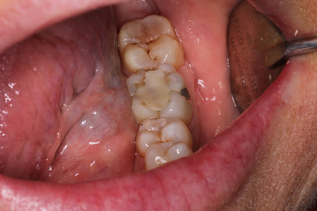 |
| Figure 1 (Initial Presentation) |
After local anaesthesia and rubber dam placement (which is important when you expect irreversible pulpitis as you may need to extirpate the pulp in the same visit: PULP TEST) I removed the old restoration. The pulp floor was stained and had an old base so I assume there was originally an amalgam that was replaced. If you look carefully you can see the crack line progressing from mesial, slightly lingually to the distal ridge where there is also a crack present (Fig 2-3). In this case in a lower molar, the lingual cusps are at the most risk of fracture as they are non working cusps and are unguarded but the opposing upper molar buccal and palatal cusp. They are likely under the highest lateral loading and as well are slightly thinner than the buccal cusps. In upper teeth the buccal cusps are at higher risk of fracture for the same reasons. Due to this I usually only cover the lingual cusps in lower molars. However in hindsight I would probably consider covering the buccal cusps for a full coverage restoration due to the fact that there is a M-D crack line and the crack looks especially sinister (It is unstained so likely formed recently after amalgam removal and hasn't had time to pick up stains from the oral environment)
 |
| Figure 2: Preparation after restoration was removed |

 |
| Figure 3: Reduction of the Lingual cusps |
 |
| Figure 4: Prep reduction guide. Notice how the reduction follows the anatomy of the occlusal surface |
In this case I cut down the lingual cusps by approximately 2mm (Figure 3 and 4) to allow enough bulk of composite resin to cover the cusp. The idea is that the physical tooth cusp is now out of occlusion and therefore the flexure that opens the crack is reduced and the crack is sealed by the restoration. In the discussion with the patient, a crown would be more ideal as it is physically encircling the tooth providing a ferrule effect that braces against fracture. In hindsight, on the lingual margin, I would do less of a butt joint and taper the prep down further to something like a shoulder. Therefore I would gain some more bracing action more similar to a full coverage crown. I would also consider taking the interproximal prep down further to the edge of the crack. On advice from an endodontist, cracks don't have to be removed in vital teeth as there is dentine tubule fluid flowing outwards that will defend against bacterial ingress into the pulp space. However in non vital teeth it is a good idea to seal against this. In this case, the enamel crack will continue to be a site of bacterial contamination which may result in recurrent caries or periodontal issues in the future.
In these cases, even with full coverage it is a fairly easy restoration to perform. I restored it is Composite resin (Figure 6). I place a Tofflemire matrix band as it requires circumferential coverage. Some difficulties could arise if the contact isn't broken but this is often the feature that allows a good contact to be present after the restoration. If the interproximal enamel margin is jagged it will stop the matrix from slipping through easier and needs to be trimmed down to a butt joint. The lingual surface will always be too bulky due to the shape of the tofflemire but this can be easily trimmed after place. I perform my bonding steps and place a layer of flowable around the edge of the Tofflemire to avoid voids at the margin. I then take a blob of packable composite and place it circumferentially around till about the level of the cusp tips. This just leaves a class 1 cavity which can be filled first buccally then lingually (Figure 5)
 |
| Figure 5: Composite building technique. Black- Tofflemire band, Blue- thin layer of flowable at edge of band, Red- Circumferential ring of composite till cusp tips, Purple- buccal cusp buildup till central fissure, Brown-Lingual cusps |
 |
| Figure 6: Final restoration |
When adjusting the occlusion, aim to have the cuspal incline in line with the adjacent molars. Too steep and the restorative cusps will take too much lateral loading and too shallow and it will be out of occlusion. It is better to be too shallow than too steep. Note in this case the significant wear facet on the 37 distobuccal and distolingual cusps. This is a classic presentation caused by a plunger cusp of an upper 7 which are generally tilted to the buccal causing the palatal cusps to dip down under the occlusal plane. In hindsight I would think about reducing this cusp to avoid similar loading on the 37 tooth. This tooth was part of my differential diagnoses but did not show any cracks on transillumination. However it is certainly at risk of this in the future. It is probably beneficial tthat the 37 is unrestored as this has likely reduced its risk of cracks in the past.
Bruxism is always a consideration in cracked tooth cases and I inevitably end up having a conversation to root out the cause of the cracks. I explain that part of the reason is there is a large restoration weakening the tooth. age is a factor as well but teeth do not usually crack for no reason. Enamel is the hardest substance in the body and generally there is a significant amount of force placed on these teeth. This may be due to occlusal factors such has the aforementioned plunger cusp but also clenching and grinding which may be stress related or due to an underlying sleep disorder. In this case the patient was slightly overweight, middle aged and short faced. She had anterior and posterior wear facets and so shows positive signs for bruxism. This is likely related to obstructive sleep apnoeas and part of her future investigations is a sleep study to root out the possible causes of her bruxism.
This is a case that should be reviewed with clinical examination and pulp tests. You may consider a PA radiograph after 2-3 months (when it is likely to show change) but it may not change your treatment. The older the patient and more restorative work the tooth has causes pulp calcification and symptoms of pulpitis may not be immediately noticeable. The first sign of pain may be pain to chew from an apical periodontitis. Ultimately, the patient should be warned that the emergence of pain is possible and is a prognostic factor in treatment. It may signify the need for further endodontic treatment or exodontia.


Comments
Post a Comment
Please leave a comment and let me know what you think or if there are any topics you would like covered in the future