Some thoughts regarding extraction mechanics and surgical planning
Molar extractions are complex things there's no doubt about it. What makes them so complex is their variation in anatomy ranging from root shape and length as well as curvature, number of roots and the increased surface area of PDL. The surrounding structures add their own complexity with variations in the density of cortical bone as well as surrounding structures of the maxillary sinus and IDN. Therefore it is prudent to plan out extractions properly and have a good idea of how the surgery will go.
Here are a few tips I've picked up on surgical planning:
-Mandibular posterior cortical bone is dense, don't expect much expansion of the socket
-Don't be afraid to raise a flap or go surgical or sectional if it will be easier. Destruction of bone is less of an issue if they aren't planning for an implant. However in the posterior region where extractions are generally more difficult, the volume of bone and soft tissue is less of an issue. Once you remove the first few mm of thin crestal bone (which would likey resorb anyway) the buccolingual dimension of bone is thicker and a thin gutter will actually preserve a fair bit of bone height if done well. It will also be less pressure on the patient's jaw and therefore less stress on the TMJ, less soft tissue and bony trauma.
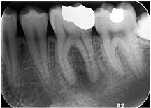
-When looking at a radiograph to plan extractions
-Follow the curve of the root, it will tell you which way the root wants to slide out. If a multi rooted teeth has roots that want to exit in different directions then the tooth must at least need to be sectioned (e.g the 36 below)
-When planning to section, compare the mid section of the crown (red line) to the mid section of the root (blue line). Usually there is a distal tilt of the roots and if you section the crown in half then the roots may split unfavourably. Instead, you must aim to section the tooth in the middle of the roots.
-Compare the width of the CEJ (Orange line) to the widest point of the roots. If the roots are wider than the CEJ it is not likely to be a simple extraction. You can expect some expansion of the bone to accommodate but not a whole lot.
-When sectioning drill down as far into the floor of the pulp chamber as possible even into the interadicular bone. This will decrease the chances of a bad split. The only place you will be wary of is an upper molar with a resorbed maxillary sinus. The xray may be misleading as the sinus can dip down between the roots to the interradicular area. Usually a bad split will be subcrestal which forces you into a surgical extraction. Don't be afraid to make the access of sectioning as wide as you need on the coronal aspect. The wider it is will ensure that the point of application of the force is as apically as possible in the base of the pulp chamber and increase the chances of a good split. Sectioning forces at the occlusal will tend to fracture the crown off.
-Root filled teeth are difficult especially with a post preparation. A 2D radiograph will given you a fair idea of where the root will fracture. Look for the area where the dentine is the thinnest from the gutta percha. Surgical extractions are very likely for root filled molars.

Here are a few tips I've picked up on surgical planning:
-Mandibular posterior cortical bone is dense, don't expect much expansion of the socket
-Don't be afraid to raise a flap or go surgical or sectional if it will be easier. Destruction of bone is less of an issue if they aren't planning for an implant. However in the posterior region where extractions are generally more difficult, the volume of bone and soft tissue is less of an issue. Once you remove the first few mm of thin crestal bone (which would likey resorb anyway) the buccolingual dimension of bone is thicker and a thin gutter will actually preserve a fair bit of bone height if done well. It will also be less pressure on the patient's jaw and therefore less stress on the TMJ, less soft tissue and bony trauma.
-When looking at a radiograph to plan extractions
-Follow the curve of the root, it will tell you which way the root wants to slide out. If a multi rooted teeth has roots that want to exit in different directions then the tooth must at least need to be sectioned (e.g the 36 below)
-When planning to section, compare the mid section of the crown (red line) to the mid section of the root (blue line). Usually there is a distal tilt of the roots and if you section the crown in half then the roots may split unfavourably. Instead, you must aim to section the tooth in the middle of the roots.
-Compare the width of the CEJ (Orange line) to the widest point of the roots. If the roots are wider than the CEJ it is not likely to be a simple extraction. You can expect some expansion of the bone to accommodate but not a whole lot.
-When sectioning drill down as far into the floor of the pulp chamber as possible even into the interadicular bone. This will decrease the chances of a bad split. The only place you will be wary of is an upper molar with a resorbed maxillary sinus. The xray may be misleading as the sinus can dip down between the roots to the interradicular area. Usually a bad split will be subcrestal which forces you into a surgical extraction. Don't be afraid to make the access of sectioning as wide as you need on the coronal aspect. The wider it is will ensure that the point of application of the force is as apically as possible in the base of the pulp chamber and increase the chances of a good split. Sectioning forces at the occlusal will tend to fracture the crown off.
-Root filled teeth are difficult especially with a post preparation. A 2D radiograph will given you a fair idea of where the root will fracture. Look for the area where the dentine is the thinnest from the gutta percha. Surgical extractions are very likely for root filled molars.
Comments
Post a Comment
Please leave a comment and let me know what you think or if there are any topics you would like covered in the future